LDHA antibody
Principal name
LDHA antibody
Alternative names for LDHA antibody
LDH-A, L-lactate dehydrogenase A chain, LDH-M, PIG19, NY-REN-59, LDH muscle subunit, Cell proliferation-inducing gene 19 protein, Renal carcinoma antigen NY-REN-59
SwissProt ID
P00338 (Human), P00339 (Pig), P00340 (Chick), P04642 (Rat), P06151 (Mouse), P13491 (Rabit), P19858 (Bovin)
Ncbi ID
NP_001075746.1, 9606, NP_005557, NP_776524, NP_001075746, NP_001158887, NP_001158887.1, NM_001135239, NM_001165414, NP_001128711, NP_034829.1, NP_034829, NM_010699, NM_017025, NM_005566, NM_001165416, NM_001165415
Available reactivities
Available hosts
Available applications
Immunocytochemistry/Immunofluorescence (ICC/IF), Immunoprecipitation (IP), Enzyme Immunoassay (E), Western blot / Immunoblot (WB), ELISA (detection) (E(detection)), Dot blot (Dot), Immunodiffusion (ID), Radioimmunoassay (R), Paraffin Sections (P), Flow Cytometry (F), Luminex (LMNX), Frozen Sections (C)
Background of LDHA antibody
Lactate dehydrogese (LDH) is a ubiquitous enzyme that catalyzes the inter-conversion of pyruvate and lactate, with simultaneous inter-conversion of DH and D+. Mammalian LDH has five tetrameric isozymes comprising combitions of two subunits (i) A subunit (LDHA or LDHM) predomites in skeletal muscle where it involve in aerobic metabolism and pyruvate reduction (ii) B subunit (LDHB) predomites in cardiac muscle where it helps catalyze aerobic oxidation of pyruvate. Several human cancers such as including rel, breast, gastric, sopharyngeal cancer etc. have higher LDHA levels compared to normal tissues and LDHA upregulation ensures efficient aerobic/glycolytic metabolism for tumor cells with reduced oxygen dependency. Moreover, LDHA also plays an important role in the development, invasion and metastasis of several maligncies and induced suppression of LDHA in cancer cells has been shown to trigger ROS burst, mitochondrial pathway apoptosis and limited tumorigenic potential. Defects in LDHA have also been linked to exertiol myoglobinuria and glycogen storage disease type 11 (GSD11).
General readings
Zhu, X., et al. Genet. Epidemiol. 34(2):171-187(2010)
Zhuang, L., et al. Mod. Pathol. 23(1):45-53(2010)
Zhao, Y.H., et al. Oncogene 28(42):3689-3701(2009)
Xie, H., et al. Mol. Cancer Ther. 8(3):626-635(2009)
Koukourakis, M.I., et al. Oncology 77(5):285-292(2009)
Rikova, K., et al. Cell 131(6):1190-1203(2007)
Rush, J., et al. Nat. Biotechnol. 23(1):94-101(2005)
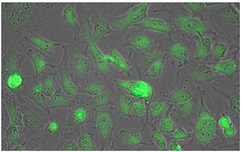
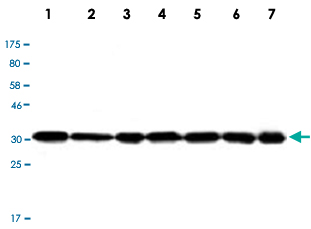
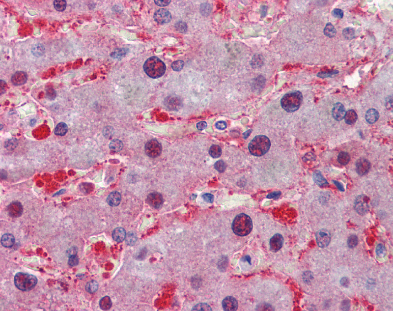
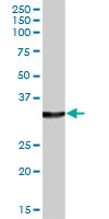
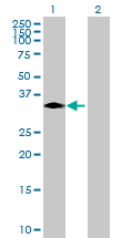
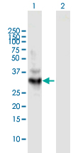
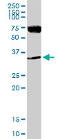
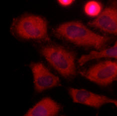
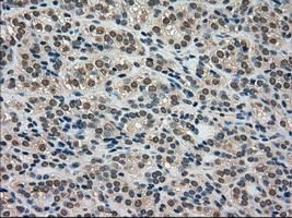
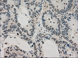
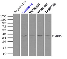
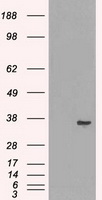
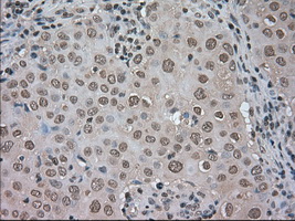
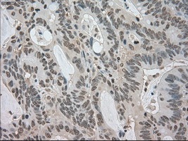
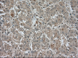
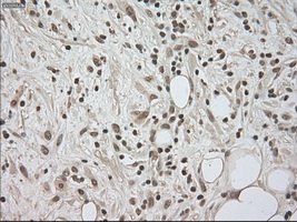
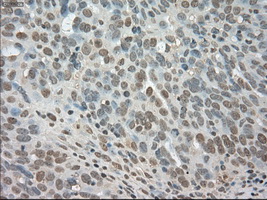
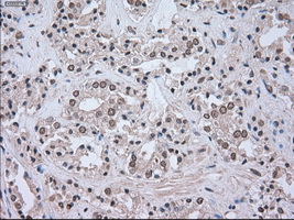
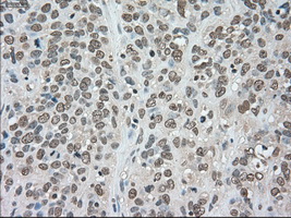
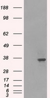
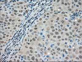
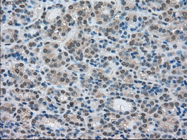
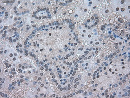
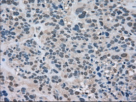
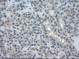
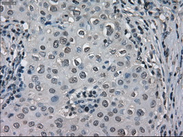
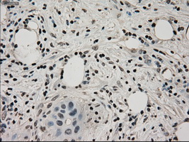
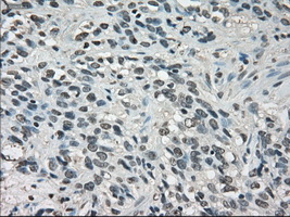
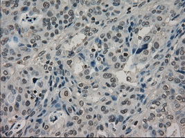
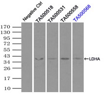
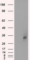
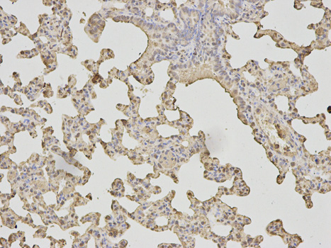
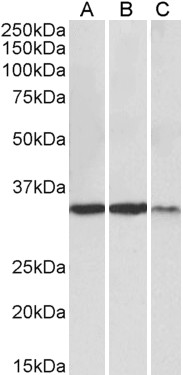
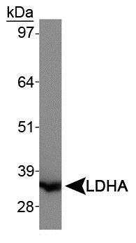
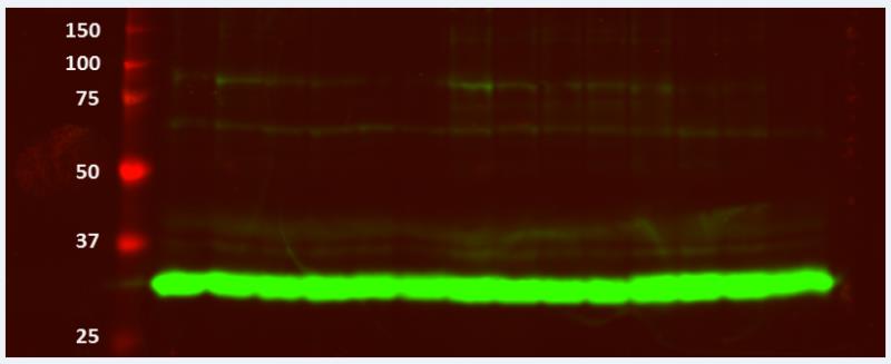
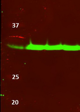
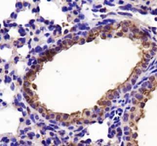
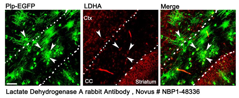
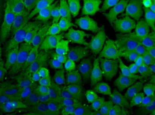
Staining: Lactate Dehydrogenase as green fluorescent signal.
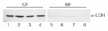
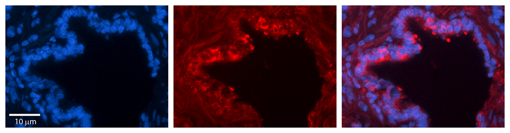
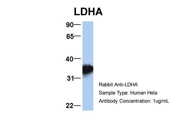
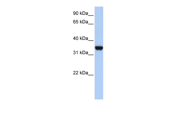
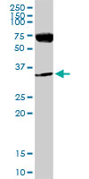
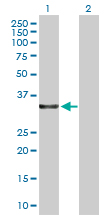
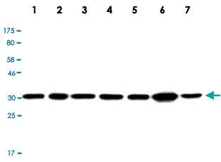


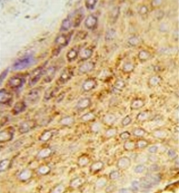
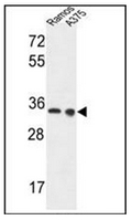
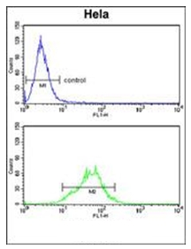
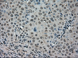













- Quick-Filters:
- Highly Validated (1)
Primary Antibodies
| Catalog No. | Host | Iso. | Clone | Pres. | React. | Applications | |
|---|---|---|---|---|---|---|---|
| TA336781 | LDHA antibody |
||||||
| Rabbit Polyclonal Lactate Dehydrogenase A/LDHA Antibody | |||||||
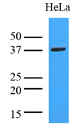
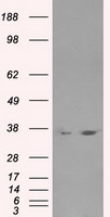
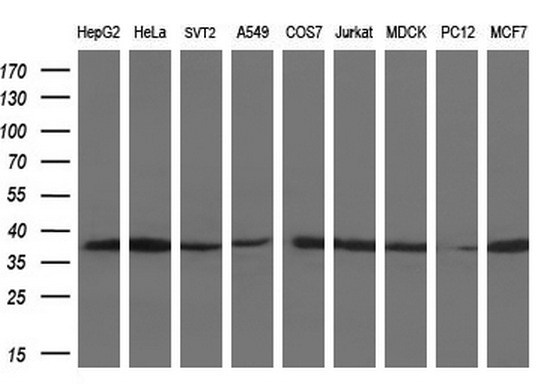
![Western Blot: Lactate Dehydrogenase A Antibody [TA336781] - Analysis of Lactate Dehydrogenase A in HeLa whole cell lysates.](https://m1.acris-antibodies.com/catalog/product/cache/2/small_image/50x50/9df78eab33525d08d6e5fb8d27136e95/OriGene/NBP1-48336-1-W.jpg) |
Rabbit | Purified | Hu, Ms | C, ICC/IF, P, WB |
0.1 ml /
$415.00
|
||
| OriGene Technologies, Inc. | |||||||
 +6 additional images +6 additional images |
Highly validated antibody! |
||||||
| TA326950 | LDHA antibody |
||||||
| Rabbit anti-LDHA Polyclonal Antibody | |||||||
 |
Rabbit | IgG | Purified | Hu, Ms, Rt |
0.1 mg /
$335.00
|
||
| OriGene Technologies, Inc. | |||||||
| +1 additional image |
|||||||
| TA327768 | LDHA (C-term) antibody |
||||||
| LDHA Goat Polyclonal Antibody | |||||||
 |
Goat | Aff - Purified | Hu | E, WB |
0.1 mg /
$360.00
|
||
| OriGene Technologies, Inc. | |||||||
| AP52459PU-N | LDHA (Center) antibody |
||||||
 |
Rabbit | Ig | Aff - Purified | Hu | F, P, WB |
0.4 ml /
$415.00
|
|
| OriGene Technologies GmbH | |||||||
| +2 additional images |
|||||||
| R1094 | LDHA antibody |
||||||
 |
Goat | Serum | Rb | E, ICC/IF, IP, WB |
2 ml /
$1,345.00
|
||
| OriGene Technologies GmbH | |||||||
| +1 additional image |
|||||||