MPP6 antibody
Principal name
MPP6 antibody
Alternative names for MPP6 antibody
VAM1, Veli-associated MAGUK 1, MAGUK p55 subfamily member 6
Ncbi ID
Available reactivities
Available hosts
Available applications
Enzyme Immunoassay (E), Western blot / Immunoblot (WB), Paraffin Sections (P), Flow Cytometry (F)
Background of MPP6 antibody
- Kit Component:
- - KN310259G1, Mpp6 gRNA vector 1 in pCas-Guide vector
- KN310259G2, Mpp6 gRNA vector 2 in pCas-Guide vector
- KN310259D, donor vector containing left and right homologous arms and Luciferase-Puro functional cassette
- GE100003, scramble sequence in pCas-Guide vector - CRISPR Manual:
- CRISPR/CAS9 Genome Editing Manual
General readings
Kamberov E, Makarova O, Roh M, Liu A, Karnak D, Straight S, Margolis B. Molecular cloning and characterization of Pals, proteins associated with mLin-7. J Biol Chem. 2000 Apr 14;275(15):11425-31. PMID: 10753959
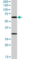
MPP6 polyclonal antibody ( Cat # PAB6552 ) staining ( 0.3 µg/ml ) of 293 lysate ( RIPA buffer, 35 µg total protein per lane ) . Primary incubated for 1 hour. Detected by western blot using chemiluminescence.
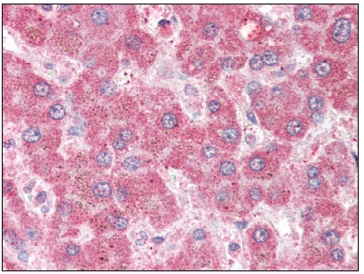
Liver: Formalin-Fixed Paraffin-Embedded (FFPE)
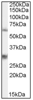
Antibody staining (0.3 µg/ml) of 293 lysate (RIPA buffer, 35 µg total protein per lane). Primary incubated for 1 hour. Detected by western blot using Chemiluminescence.
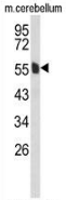
Western blot analysis of MPP6 (arrow) in mouse cerebellum tissue lysates (35ug/lane) using MPP6 antibody Cat.-No. AP52741PU-N.

Flow cytometric analysis of 293 cells (right histogram) compared to a negative control cell (left histogram) using MPP6 antibody Cat.-No. AP52741PU-N, followed by FITC-conjugated goat-anti-rabbit secondary antibodies.

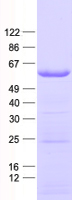
HEK293T cells were transfected with the pCMV6-ENTRY control (Left lane) or pCMV6-ENTRY MPP6 (RC205667, Right lane) cDNA for 48 hrs and lysed. Equivalent amounts of cell lysates (5 ug per lane) were separated by SDS-PAGE and immunoblotted with anti-MPP6.(1:2000)
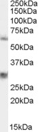
TA302912 staining (0.3µg/ml) of 293 lysate (RIPA buffer, 35µg total protein per lane). Primary incubated for 1 hour. Detected by western blot using chemiluminescence.
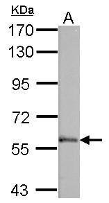
Sample (50 ug of whole cell lysate). A: mouse brain. 7.5% SDS PAGE. TA308355 diluted at 1:1000.
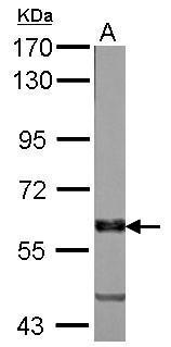
Sample (50 ug of whole cell lysate). A: Rat brain. 7.5% SDS PAGE. TA308355 diluted at 1:1000.
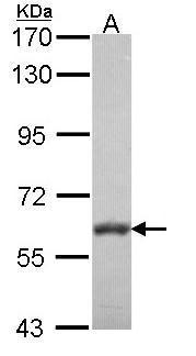
Sample (30 ug of whole cell lysate). A: NT2D1. 7.5% SDS PAGE. TA308355 diluted at 1:1000.
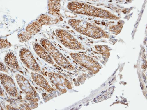
Immunohistochemical analysis of paraffin-embedded Colon, using VAM1(TA308355) antibody at 1:100 dilution.
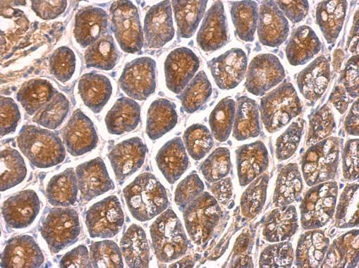
VAM1 antibody [N1], N-term detects VAM1 protein at on mouse intestine by immunohistochemical analysis. Sample: Paraffin-embedded mouse intestine. VAM1 antibody [N1], N-term (TA308355) dilution: 1:500.
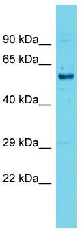
Host: Rabbit; Target Name: Mpp6; Sample Tissue: Mouse Testis lysates; Antibody Dilution: 1.0 ug/ml
- Quick-Filters:
- Highly Validated (remove)
Primary Antibodies
| Catalog No. | Host | Iso. | Clone | Pres. | React. | Applications | |
|---|---|---|---|---|---|---|---|
| TA308355 | MPP6 antibody |
||||||
| Rabbit Polyclonal antibody to VAM1 (membrane protein, palmitoylated 6 (MAGUK p55 subfamily member 6)) | |||||||
 |
Rabbit | IgG | Aff - Purified | Hu, Ms, Rt | P, WB |
0.1 ml /
$415.00
|
|
| OriGene Technologies, Inc. | |||||||
 +4 additional images +4 additional images |
Highly validated antibody! |
||||||