EGR1 antibody
Principal name
EGR1 antibody
Alternative names for EGR1 antibody
Early growth response protein 1, EGR-1, Zif268, Zif-268, NGFI-A, AT225, ETR103, KROX-24, Protein Krox-24, Transcription factor Zif268, Nerve growth factor-induced protein A, Transcription factor ETR103
Ncbi ID
9606, NP_031939, NP_001955.1, NP_001955, NM_007913, ACE87567.1, NP_031939.1, NM_001964
Available reactivities
Available hosts
Available applications
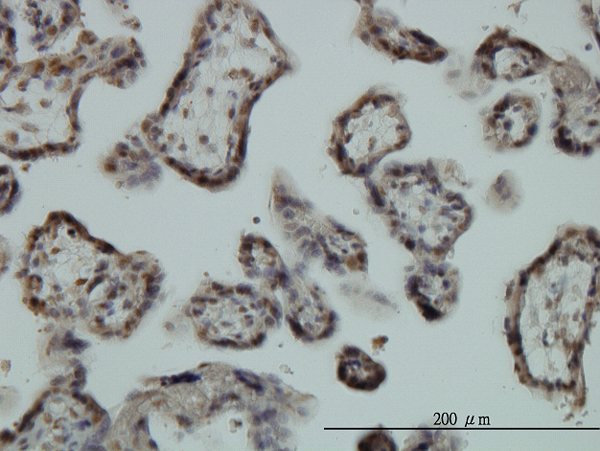
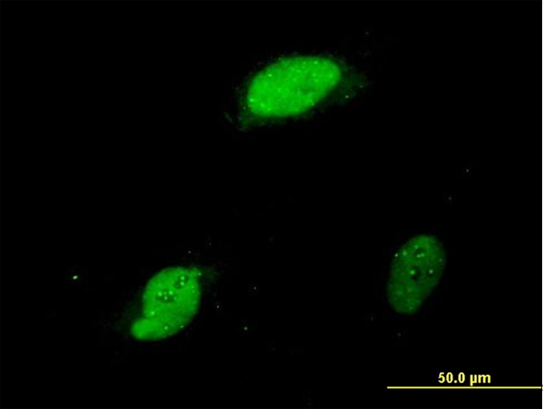
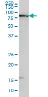
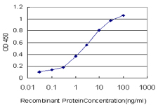
Enzyme Immunoassay (E), Western blot / Immunoblot (WB), Immunocytochemistry/Immunofluorescence (ICC/IF), Paraffin Sections (P)
Background of EGR1 antibody
Egr proteins function in transcription regulatory activities surrounding cellular growth, differentiation and function. The deduced amino acid sequences of human Egr-2 and mouse Egr-1 are 92% identical in the zinc finger region but show no homology elsewhere. Egr-2 is a sequence-specific DNA-binding transcription factor that binds two specific DNA sites located in the promoter region of HoxA4 and localizes to the nucleus. Defects in the Egr-2 protein are a cause of congenital hypomyelination neuropathy (CHN). CHN is characterized clinically by early onset of hypotonia, areflexia, distal muscle weakness and very slow nerve conduction velocities. Mutations in the gene that encodes Egr-2 (EGR2) also cause of Dejerine-Sottas syndrome (DSS), which is also known as Dejerine-Sottas neuropathy (DSN) or hereditary motor and sensory neuropathy III (HMSN3). DSS patients exhibit severe early onset motor and sensory neuropathy with very slow nerve conduction velocities and elevated cerebrospinal fluid protein concentrations.
General readings
Schnell,F.J., et al., (2006) J. Immunol. 176 (8), 4740-4747

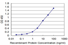
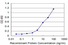
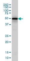
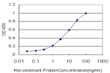
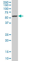
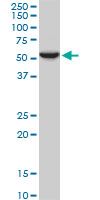
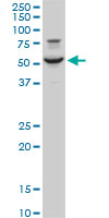
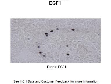






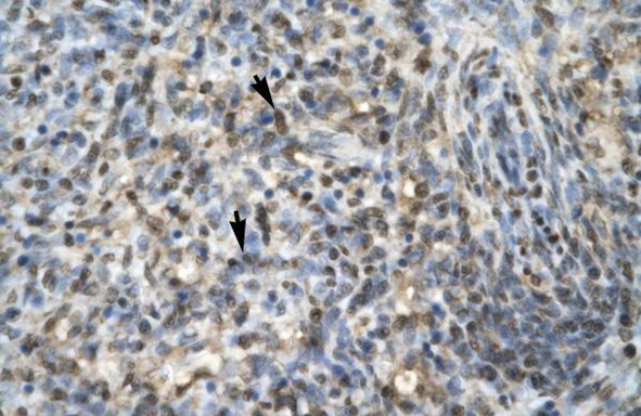
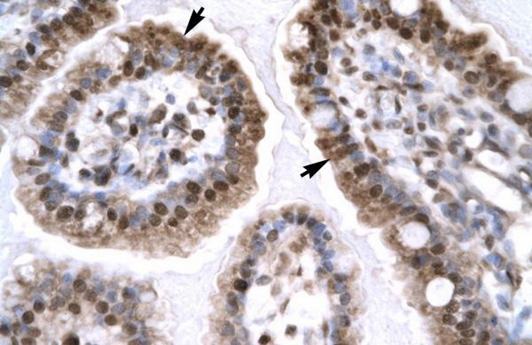
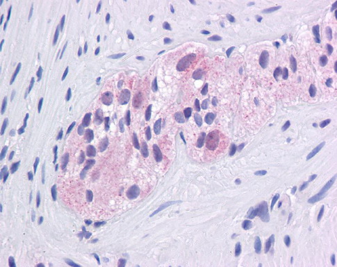

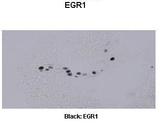
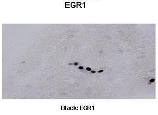
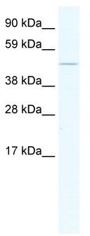
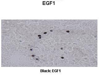
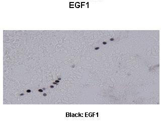
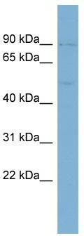
Primary Antibodies
| Catalog No. | Host | Iso. | Clone | Pres. | React. | Applications | |
|---|---|---|---|---|---|---|---|
| TA319423 | EGR1 (94-108) antibody |
||||||
| Rabbit polyclonal anti-EGR-1 antibody | |||||||
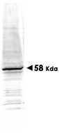
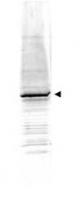
![WB using Anti-EGR-1 antibody shows detection of a predominant band at ~58 kDa corresponding to EGR-1 present in mouse embryonic fibroblast whole cell lysate (arrowhead). Primary antibody was used at a 1,500 dilution. Reaction occurred 2h at room temperature followed by washes and reaction with a 1:10,000 dilution of IRDye™800 conjugated Gt-a-Rabbit IgG [H&L] MX for 45 min at room temperature.](https://m1.acris-antibodies.com/catalog/product/cache/2/small_image/50x50/9df78eab33525d08d6e5fb8d27136e95/OriGene/600-401-693-1-W.jpg) |
Rabbit | IgG | Aff - Purified | Chimp, Hu, Ms, Rt | E, P, WB |
0.1 mg /
$515.00
|
|
| OriGene Technologies, Inc. | |||||||
 +1 additional image +1 additional image |
|||||||