DOK7 antibody
Principal name
DOK7 antibody
Alternative names for DOK7 antibody
C4orf25, Downstream of tyrosine kinase 7
Ncbi ID
9606, NM_173660, NP_001158145.1, NP_775931, NP_766296.1, NM_172708
Available reactivities
Available hosts
Available applications
Enzyme Immunoassay (E), Immunocytochemistry/Immunofluorescence (ICC/IF), Paraffin Sections (P), Western blot / Immunoblot (WB), Flow Cytometry (F)
Background of DOK7 antibody
- Kit Component:
- - KN304752G1, Dok7 gRNA vector 1 in pCas-Guide vector
- KN304752G2, Dok7 gRNA vector 2 in pCas-Guide vector
- KN304752D, donor vector containing left and right homologous arms and Luciferase-Puro functional cassette
- GE100003, scramble sequence in pCas-Guide vector - CRISPR Manual:
- CRISPR/CAS9 Genome Editing Manual


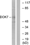
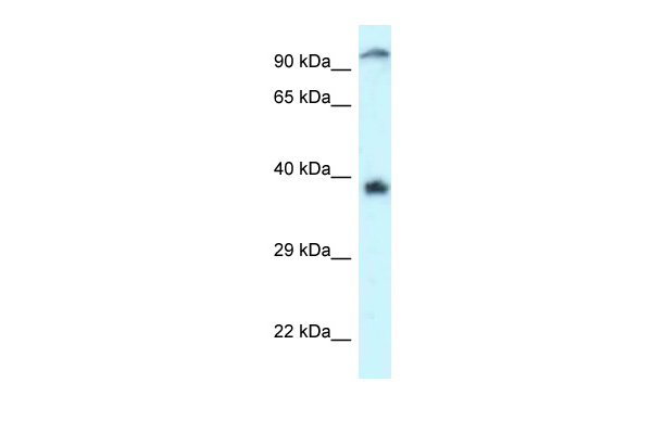
Human Jurkat; WB Suggested Anti-DOK7 Antibody. . Titration: 1.0 ug/ml. . Positive Control: Jurkat Whole Cell; DOK7 antibody - C-terminal region (ARP61801_P050) in Human Jurkat cells using Western Blot
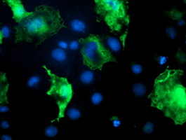
Anti-DOK7 mouse monoclonal antibody (TA504786) immunofluorescent staining of COS7 cells transiently transfected by pCMV6-ENTRY DOK7(RC219267).
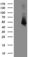
HEK293T cells were transfected with the pCMV6-ENTRY control (Left lane) or pCMV6-ENTRY DOK7 (RC219267, Right lane) cDNA for 48 hrs and lysed. Equivalent amounts of cell lysates (5 ug per lane) were separated by SDS-PAGE and immunoblotted with anti-DOK7.
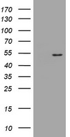
HEK293T cells were transfected with the pCMV6-ENTRY control (Left lane) or pCMV6-ENTRY DOK7 (RC219267, Right lane) cDNA for 48 hrs and lysed. Equivalent amounts of cell lysates (5 ug per lane) were separated by SDS-PAGE and immunoblotted with anti-DOK7.

Immunohistochemical staining of paraffin-embedded Human Kidney tissue within the normal limits using anti-DOK7 mouse monoclonal antibody. (Heat-induced epitope retrieval by 10mM citric buffer, pH6.0, 100C for 10min, TA504788)
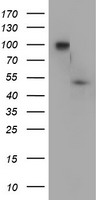
HEK293T cells were transfected with the pCMV6-ENTRY control (Left lane) or pCMV6-ENTRY DOK7 (RC219267, Right lane) cDNA for 48 hrs and lysed. Equivalent amounts of cell lysates (5 ug per lane) were separated by SDS-PAGE and immunoblotted with anti-DOK7.
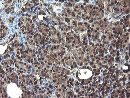
Immunohistochemical staining of paraffin-embedded Human pancreas tissue within the normal limits using anti-DOK7 mouse monoclonal antibody. (Heat-induced epitope retrieval by 10mM citric buffer, pH6.0, 100C for 10min, TA504789)
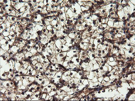
Immunohistochemical staining of paraffin-embedded Carcinoma of Human kidney tissue using anti-DOK7 mouse monoclonal antibody. (Heat-induced epitope retrieval by 10mM citric buffer, pH6.0, 100C for 10min, TA504789)

Immunohistochemical staining of paraffin-embedded Adenocarcinoma of Human ovary tissue using anti-DOK7 mouse monoclonal antibody. (Heat-induced epitope retrieval by 10mM citric buffer, pH6.0, 100C for 10min, TA504789)
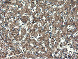
Immunohistochemical staining of paraffin-embedded Human liver tissue within the normal limits using anti-DOK7 mouse monoclonal antibody. (Heat-induced epitope retrieval by 10mM citric buffer, pH6.0, 100C for 10min, TA504789)
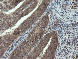
Immunohistochemical staining of paraffin-embedded Adenocarcinoma of Human endometrium tissue using anti-DOK7 mouse monoclonal antibody. (Heat-induced epitope retrieval by 10mM citric buffer, pH6.0, 100C for 10min, TA504789)
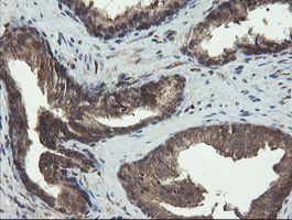
Immunohistochemical staining of paraffin-embedded Human prostate tissue within the normal limits using anti-DOK7 mouse monoclonal antibody. (Heat-induced epitope retrieval by 10mM citric buffer, pH6.0, 100C for 10min, TA504789)
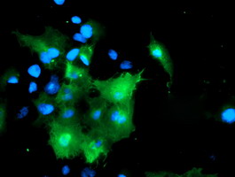
Anti-DOK7 mouse monoclonal antibody (TA504789) immunofluorescent staining of COS7 cells transiently transfected by pCMV6-ENTRY DOK7(RC219267).
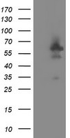
HEK293T cells were transfected with the pCMV6-ENTRY control (Left lane) or pCMV6-ENTRY DOK7 (RC219267, Right lane) cDNA for 48 hrs and lysed. Equivalent amounts of cell lysates (5 ug per lane) were separated by SDS-PAGE and immunoblotted with anti-DOK7.

HEK293T cells transfected with either RC219267 overexpress plasmid(Red) or empty vector control plasmid(Blue) were immunostained by anti-DOK7 antibody(TA504789), and then analyzed by flow cytometry.
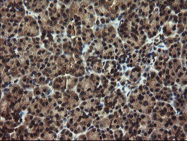
Immunohistochemical staining of paraffin-embedded Human pancreas tissue within the normal limits using anti-DOK7 mouse monoclonal antibody. (Heat-induced epitope retrieval by 10mM citric buffer, pH6.0, 100C for 10min, TA504801)

Immunohistochemical staining of paraffin-embedded Adenocarcinoma of Human ovary tissue using anti-DOK7 mouse monoclonal antibody. (Heat-induced epitope retrieval by 10mM citric buffer, pH6.0, 100C for 10min, TA504801)
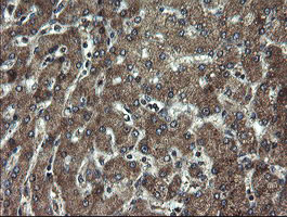
Immunohistochemical staining of paraffin-embedded Human liver tissue within the normal limits using anti-DOK7 mouse monoclonal antibody. (Heat-induced epitope retrieval by 10mM citric buffer, pH6.0, 100C for 10min, TA504801)

Immunohistochemical staining of paraffin-embedded Adenocarcinoma of Human endometrium tissue using anti-DOK7 mouse monoclonal antibody. (Heat-induced epitope retrieval by 10mM citric buffer, pH6.0, 100C for 10min, TA504801)
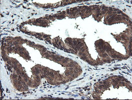
Immunohistochemical staining of paraffin-embedded Human prostate tissue within the normal limits using anti-DOK7 mouse monoclonal antibody. (Heat-induced epitope retrieval by 10mM citric buffer, pH6.0, 100C for 10min, TA504801)
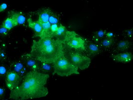
Anti-DOK7 mouse monoclonal antibody (TA504801) immunofluorescent staining of COS7 cells transiently transfected by pCMV6-ENTRY DOK7(RC219267).
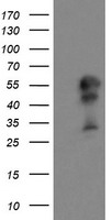
HEK293T cells were transfected with the pCMV6-ENTRY control (Left lane) or pCMV6-ENTRY DOK7 (RC219267, Right lane) cDNA for 48 hrs and lysed. Equivalent amounts of cell lysates (5 ug per lane) were separated by SDS-PAGE and immunoblotted with anti-DOK7.

Western blot analysis of extracts (35ug) from 9 different cell lines by usin g anti-DOK7 monoclonal antibody (HepG2: human; HeLa: human; SVT2: mouse; A549: human; COS7: monkey; Jurkat: human; MDCK: canine; PC12: rat; MCF7: human).
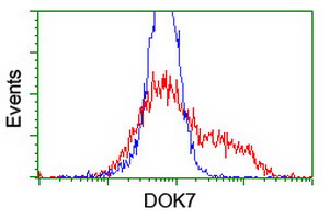
HEK293T cells transfected with either RC219267 overexpress plasmid(Red) or empty vector control plasmid(Blue) were immunostained by anti-DOK7 antibody(TA504801), and then analyzed by flow cytometry.
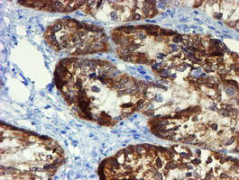
Immunohistochemical staining of paraffin-embedded Adenocarcinoma of Human ovary tissue using anti-DOK7 mouse monoclonal antibody. (Heat-induced epitope retrieval by 10mM citric buffer, pH6.0, 100C for 10min, TA504809)
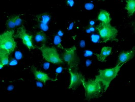
Anti-DOK7 mouse monoclonal antibody (TA504809) immunofluorescent staining of COS7 cells transiently transfected by pCMV6-ENTRY DOK7(RC219267).
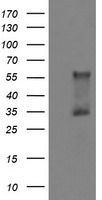
HEK293T cells were transfected with the pCMV6-ENTRY control (Left lane) or pCMV6-ENTRY DOK7 (RC219267, Right lane) cDNA for 48 hrs and lysed. Equivalent amounts of cell lysates (5 ug per lane) were separated by SDS-PAGE and immunoblotted with anti-DOK7.
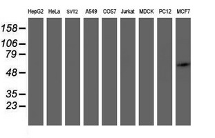
Western blot analysis of extracts (35ug) from 9 different cell lines by usin g anti-DOK7 monoclonal antibody (HepG2: human; HeLa: human; SVT2: mouse; A549: human; COS7: monkey; Jurkat: human; MDCK: canine; PC12: rat; MCF7: human).
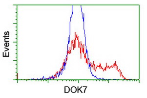
HEK293T cells transfected with either RC219267 overexpress plasmid(Red) or empty vector control plasmid(Blue) were immunostained by anti-DOK7 antibody(TA504809), and then analyzed by flow cytometry.
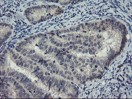
Immunohistochemical staining of paraffin-embedded Adenocarcinoma of Human endometrium tissue using anti-DOK7 mouse monoclonal antibody. (Heat-induced epitope retrieval by 10mM citric buffer, pH6.0, 100C for 10min, TA504815)
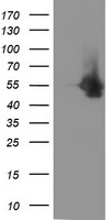
HEK293T cells were transfected with the pCMV6-ENTRY control (Left lane) or pCMV6-ENTRY DOK7 (RC219267, Right lane) cDNA for 48 hrs and lysed. Equivalent amounts of cell lysates (5 ug per lane) were separated by SDS-PAGE and immunoblotted with anti-DOK7.
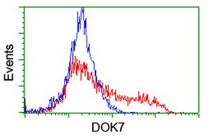
HEK293T cells transfected with either RC219267 overexpress plasmid(Red) or empty vector control plasmid(Blue) were immunostained by anti-DOK7 antibody(TA504815), and then analyzed by flow cytometry.
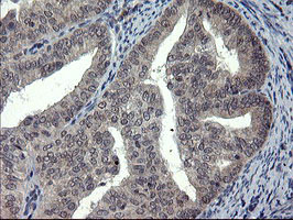
Immunohistochemical staining of paraffin-embedded Adenocarcinoma of Human endometrium tissue using anti-DOK7 mouse monoclonal antibody. (Heat-induced epitope retrieval by 10mM citric buffer, pH6.0, 100C for 10min, TA504816)
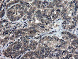
Immunohistochemical staining of paraffin-embedded Adenocarcinoma of Human ovary tissue using anti-DOK7 mouse monoclonal antibody. (Heat-induced epitope retrieval by 10mM citric buffer, pH6.0, 100C for 10min, TA504816)

Immunohistochemical staining of paraffin-embedded Carcinoma of Human thyroid tissue using anti-DOK7 mouse monoclonal antibody. (Heat-induced epitope retrieval by 10mM citric buffer, pH6.0, 100C for 10min, TA504816)
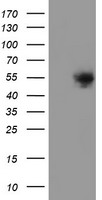
HEK293T cells were transfected with the pCMV6-ENTRY control (Left lane) or pCMV6-ENTRY DOK7 (RC219267, Right lane) cDNA for 48 hrs and lysed. Equivalent amounts of cell lysates (5 ug per lane) were separated by SDS-PAGE and immunoblotted with anti-DOK7.
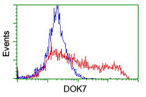
HEK293T cells transfected with either RC219267 overexpress plasmid(Red) or empty vector control plasmid(Blue) were immunostained by anti-DOK7 antibody(TA504816), and then analyzed by flow cytometry.
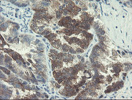
Immunohistochemical staining of paraffin-embedded Adenocarcinoma of Human ovary tissue using anti-DOK7 mouse monoclonal antibody. (Heat-induced epitope retrieval by 10mM citric buffer, pH6.0, 100C for 10min, TA504825)
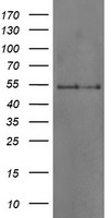
HEK293T cells were transfected with the pCMV6-ENTRY control (Left lane) or pCMV6-ENTRY DOK7 (RC219267, Right lane) cDNA for 48 hrs and lysed. Equivalent amounts of cell lysates (5 ug per lane) were separated by SDS-PAGE and immunoblotted with anti-DOK7.
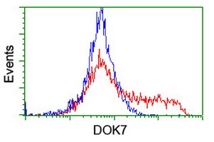
HEK293T cells transfected with either RC219267 overexpress plasmid(Red) or empty vector control plasmid(Blue) were immunostained by anti-DOK7 antibody(TA504825), and then analyzed by flow cytometry.
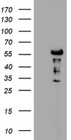
HEK293T cells were transfected with the pCMV6-ENTRY control (Left lane) or pCMV6-ENTRY DOK7 (RC219267, Right lane) cDNA for 48 hrs and lysed. Equivalent amounts of cell lysates (5 ug per lane) were separated by SDS-PAGE and immunoblotted with anti-DOK7.
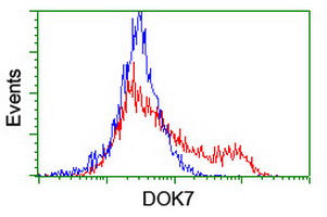
HEK293T cells transfected with either RC219267 overexpress plasmid(Red) or empty vector control plasmid(Blue) were immunostained by anti-DOK7 antibody(TA504832), and then analyzed by flow cytometry.

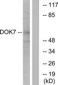
Western blot analysis of extracts from mouse brain cells, using DOK7 antibody.The lane on the right is treated with the synthesized peptide.
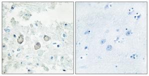
Immunohistochemistry analysis of paraffin-embedded human brain tissue using DOK7 antibody.The picture on the right is treated with the synthesized peptide.
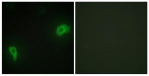
Immunofluorescence analysis of HepG2 cells, using DOK7 antibody.The picture on the right is treated with the synthesized peptide.
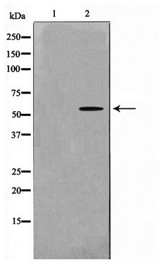
Western blot analysis on mouse brain cell lysate using DOK7 Antibody
- Quick-Filters:
- Highly Validated (3)
Primary Antibodies
| Catalog No. | Host | Iso. | Clone | Pres. | React. | Applications | |
|---|---|---|---|---|---|---|---|
| TA504789 | DOK7 antibody |
||||||
| DOK7 mouse monoclonal antibody, clone OTI1F7 (formerly 1F7) | |||||||
 |
Mouse | IgG1 | OTI1F7 | Purified | Hu | F, ICC/IF, P, WB |
0.1 ml /
$360.00
|
| OriGene Technologies, Inc. | |||||||
 +8 additional images +8 additional images |
Highly validated antibody!
|
||||||
| TA504801 | DOK7 antibody |
||||||
| DOK7 mouse monoclonal antibody, clone OTI1A9 (formerly 1A9) | |||||||
 |
Mouse | IgG1 | OTI1A9 | Purified | Hu, Ms | F, ICC/IF, P, WB |
0.1 ml /
$379.00
|
| OriGene Technologies, Inc. | |||||||
| +8 additional images |
Highly validated antibody!
|
||||||
| TA504809 | DOK7 antibody |
||||||
| DOK7 mouse monoclonal antibody, clone OTI5G8 (formerly 5G8) | |||||||
 |
Mouse | IgG1 | OTI5G8 | Purified | Hu | F, ICC/IF, P, WB |
0.1 ml /
$360.00
|
| OriGene Technologies, Inc. | |||||||
| +4 additional images |
Highly validated antibody!
|
||||||
| TA312563 | DOK7 antibody |
||||||
| Rabbit polyclonal anti-DOK7 antibody | |||||||
 |
Rabbit | IgG | Purified | Hu, Ms | ICC/IF, WB |
0.1 ml /
$335.00
|
|
| OriGene Technologies, Inc. | |||||||
| +2 additional images |
|||||||
| TA504786 | DOK7 antibody |
||||||
| DOK7 mouse monoclonal antibody, clone OTI4C6 (formerly 4C6) | |||||||
 |
Mouse | IgG1 | OTI4C6 | Purified | Hu | ICC/IF, WB |
0.1 ml /
$360.00
|
| OriGene Technologies, Inc. | |||||||
| +1 additional image |
|
||||||