CAMK2A antibody
Principal name
CAMK2A antibody
Alternative names for CAMK2A antibody
CAMKA, KIAA0968, CaM-kinase II alpha chain, CaM kinase II subunit alpha, CaMK-II subunit alpha, Calcium/calmodulin-dependent protein kinase type II alpha chain
Ncbi ID
NP_741960.1, NP_741960, NP_037052.1, 9606, NP_037052, NM_171825, NP_057065.2, NP_033922.1, NM_177407, NM_009792, NM_012920, NM_015981
Available reactivities
Available hosts
Available applications
Western blot / Immunoblot (WB), Frozen Sections (C), Enzyme Immunoassay (E), Immunocytochemistry/Immunofluorescence (ICC/IF), ELISA (detection) (E(detection)), Dot blot (Dot), Paraffin Sections (P), Immunoprecipitation (IP), Radioimmunoassay (R)
Background of CAMK2A antibody
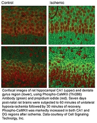
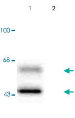
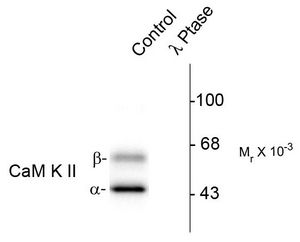
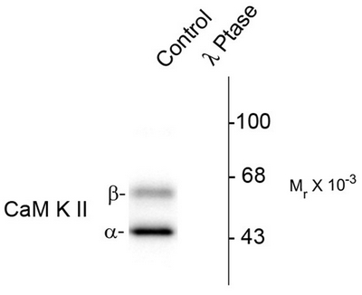
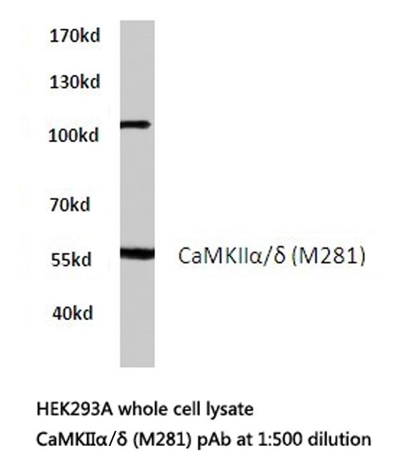
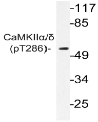
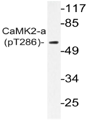
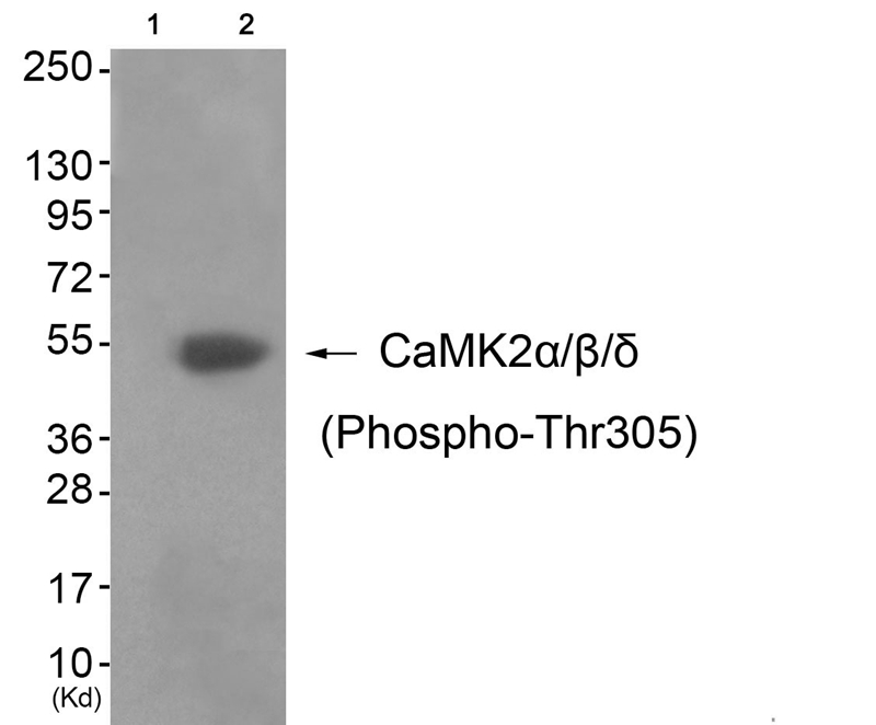
Ca2+/CalmodulinDependent Protein Kinase II (CaM Kinase II) is a multifunctional calcium and calmodulindependent protein kinase that mediates cellular responses to a wide variety of intercellular signals. CaM Kinase II has been shown to regulate diverse cellular functions including synaptic plasticity, neurotransmitter synthesis and release, gene expression, ion channel function, carbohydrate metabolism, cytoskeletal function, and Ca2+homeostasis. Phosphorylation of Thr286 on the kinase produces an autonomously active form of CaM Kinase II. Autophosphorylation of Thr305 inhibits the activity CaM Kinase II. Phosphorylation at this site appears to reduce the association of CaM Kinase II with the PSD and reduce LTP and learning.
CaM kinase II, in a calcium and calmodulin dependent manner, phosphorylates many different brain substrates including synapsin I, tryptophan hydroxylase, tyrosine hydroxylase and nitric oxide synthase thereby performing regulatory functions associated with increases in intracellular free calcium. CaM kinase II is particularly abundant in the hippocampus and forebrain where it comprises ~1% of total protein. Within the neuron, CaM kinase II is a major component of the postsynaptic density fraction accounting for as much as 30% of total protein in the post synaptic density (PSD) region. Autophosphorylation of threonine-286 occurs after calcium/calmodulin activation and can enable CaM kinase II to remain active independent of free calcium. Autophosphorylation of CaM kinase II in hippocampal neurons, which results in relatively high levels of kinase activity even at basal calcium concentrations, may allow for both upward and downward regulation as opposed to simply on/off regulation.
General readings
J Bossuyt, K Helmstadter, X Wu, et al. (2008) Ca2+/Calmodulin-Dependent Protein Kinase II {delta} and Protein Kinase D Overexpression Reinforce. Circulation Research, 102(6):695-702.

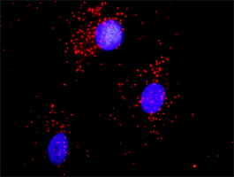
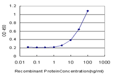
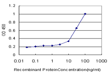
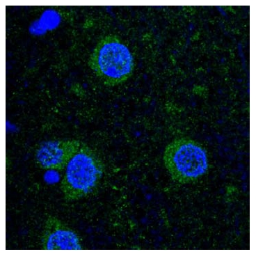

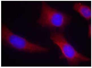

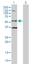
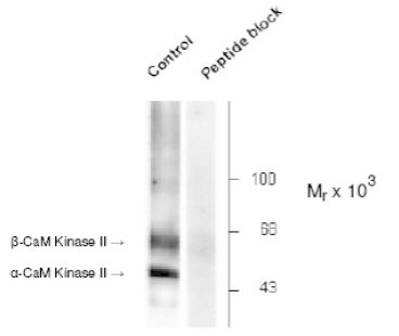
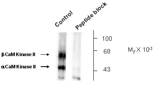
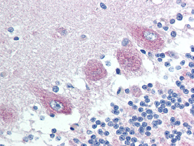
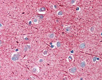
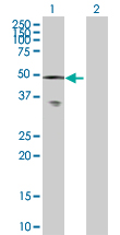
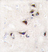
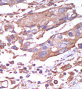
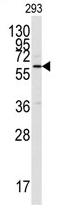
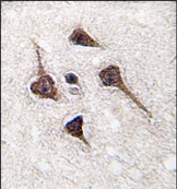
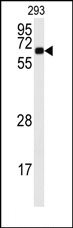
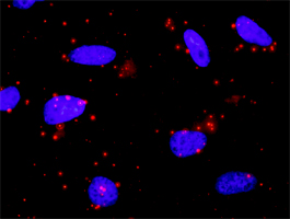
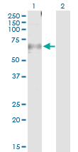


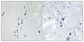
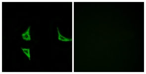
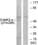
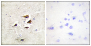
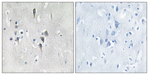
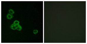
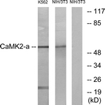
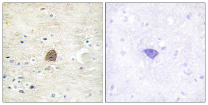

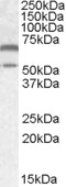
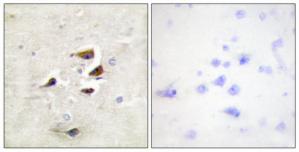
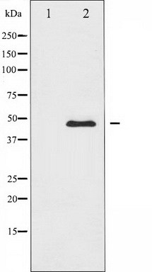


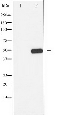


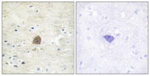

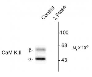
Primary Antibodies
| Catalog No. | Host | Iso. | Clone | Pres. | React. | Applications | |
|---|---|---|---|---|---|---|---|
| TA313546 | CAMK2A antibody |
||||||
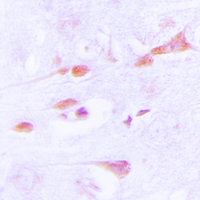
| Rabbit polyclonal CaMKII (Thr305) antibody(Phospho-specific) | |||||||
 |
Rabbit | IgG | Purified | Hu, Ms, Rt | E, P, WB |
0.1 ml /
$335.00
|
|
| OriGene Technologies, Inc. | |||||||
 +2 additional images +2 additional images |
|||||||
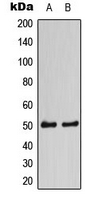
| AP13884PU-N | CAMK2A (C-term) antibody |
||||||
 |
Rabbit | Ig | Purified | Hu | P, WB |
0.4 ml /
$415.00
|
|
| OriGene Technologies GmbH | |||||||
| +2 additional images |
|||||||