BLK antibody
Principal name
BLK antibody
Alternative names for BLK antibody
B lymphocyte kinase, p55-BLK, p55BLK, Tyrosine-protein kinase BLK
Ncbi ID
NP_001706.2, 9606, NP_001706, BC007371, NM_001715.2, NM_007549, NM_001715
Available reactivities
Available hosts
Available applications
Western blot / Immunoblot (WB), Enzyme Immunoassay (E), Paraffin Sections (P), ELISA (detection) (E(detection)), Immunoprecipitation (IP), Flow Cytometry (F), Immunocytochemistry/Immunofluorescence (ICC/IF)
Background of BLK antibody
Protein kinases are enzymes that transfer a phosphate group from a phosphate donor, generally the g phosphate of ATP, onto an acceptor amino acid in a substrate protein. By this basic mechanism, protein kinases mediate most of the signal transduction in eukaryotic cells, regulating cellular metabolism, transcription, cell cycle progression, cytoskeletal rearrangement and cell movement, apoptosis, and differentiation. With more than 500 gene products, the protein kinase family is one of the largest families of proteins in eukaryotes. The family has been classified in 8 major groups based on sequence comparison of their tyrosine (PTK) or serine/threonine (STK) kinase catalytic domains. The STE group (homologs of yeast Sterile 7, 11, 20 kinases) consists of 50 kinases related to the mitogen-activated protein kinase (MAPK) cascade families (Ste7/MAP2K, Ste11/MAP3K, and Ste20/MAP4K). MAP kinase cascades, consisting of a MAPK and one or more upstream regulatory kinases (MAPKKs) have been best characterized in the yeast pheromone response pathway. Pheromones bind to Ste cell surface receptors and activate yeast MAPK pathway.
General readings
Dymecki. et al. (1990) Science. 247:332-6.Tatosyan, A.G. et al. (2000) Biochemistry. 65:49-58
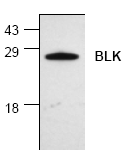
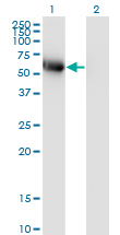
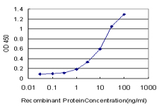
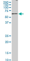
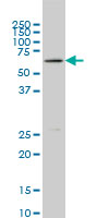
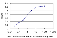
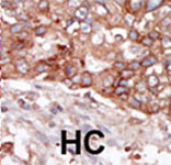
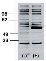
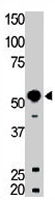
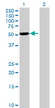
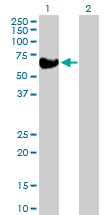
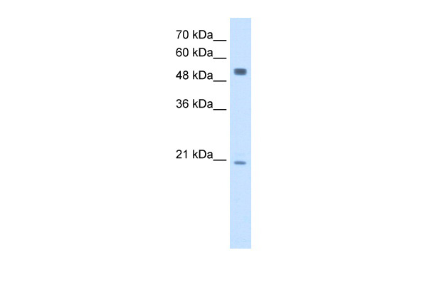
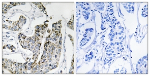
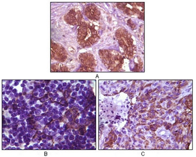

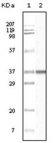
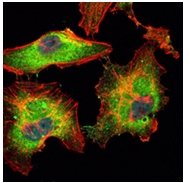
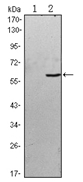


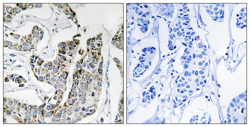
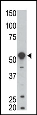
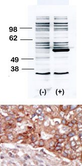
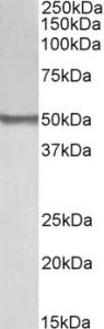
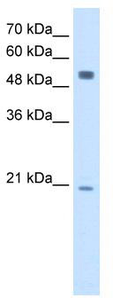

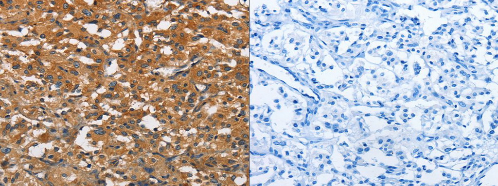
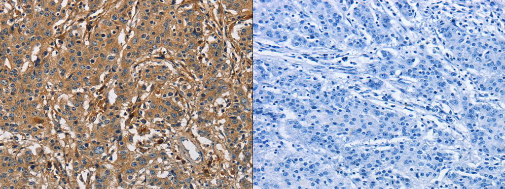
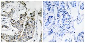
Primary Antibodies
| Catalog No. | Host | Iso. | Clone | Pres. | React. | Applications | |
|---|---|---|---|---|---|---|---|
| AM06135SU-N | BLK antibody |
||||||
 |
Mouse | IgG2a | 9D10D1 | Ascites | Hu | P |
0.1 ml /
$420.00
|
| OriGene Technologies GmbH | |||||||
| AM06138SU-N | BLK antibody |
||||||
 |
Mouse | IgG1 | 9D10B7H6 | Ascites | Hu | E, P, WB |
0.1 ml /
$340.00
|
| OriGene Technologies GmbH | |||||||
 +1 additional image +1 additional image |
|||||||
| AM06549SU-N | BLK antibody |
||||||
 |
Mouse | IgG1 | 1E6 | Ascites | Hu | E, F, ICC/IF, WB |
0.1 ml /
$380.00
|
| OriGene Technologies GmbH | |||||||
| +2 additional images |
|||||||