ANKRD26 antibody
Principal name
ANKRD26 antibody
Alternative names for ANKRD26 antibody
KIAA1074, Ankyrin repeat domain-containing protein 26
Ncbi ID
Available reactivities
Available hosts
Available applications
Enzyme Immunoassay (E), Western blot / Immunoblot (WB), Paraffin Sections (P)
Background of ANKRD26 antibody
- Kit Component:
- - KN216936G1, ANKRD26 gRNA vector 1 in pCas-Guide vector
- KN216936G2, ANKRD26 gRNA vector 2 in pCas-Guide vector
- KN216936D, donor vector containing left and right homologous arms and Luciferase-Puro functional cassette
- GE100003, scramble sequence in pCas-Guide vector - CRISPR Manual:
- CRISPR/CAS9 Genome Editing Manual
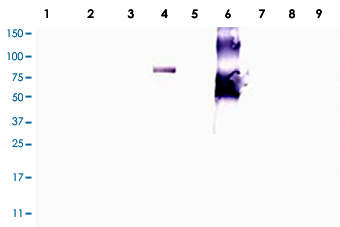
Western blot using Ankrd26 polyclonal antibody ( Cat # PAB10090 ) shows detection of a band at ~81 kDa corresponding to mouse Ankrd26 protein.Lane 1 Blank, Lane 2 MES cell lysate - 80 µg, Lane 3MES cell lysate-40 µg, Lane 4 293T-Ankrd26 transfected cell lysate-20 µg, Lane 5 control 293T celllysate-20 µg, Lane 6 BSA-Ankrd26 conjugate-20ng, Lane 7 BSA-500 ng, Lane 8 BSA-100 ng, Lane 9 BSA-20 ng.Detection of endogenous Ankrd26 protein in MES cell lysates may occur when detection methods with higher sensitivity are used.Proteins were separated by SDS-PAGE, transferred to nitrocellulose, and probed withthe primary antibody diluted to 1:1,000 followed by detection using ALP conjugated Gt-a-Rabbit IgG diluted to 1:3,000.Sizeestimation was made by comparison to prestained MW markers as indicated.Personal Communication.Ira Pastan, NIH, CCR, Bethesda, MD.

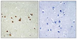
Immunohistochemistry analysis of paraffin-embedded human brain tissue using ANKRD26 antibody.The picture on the right is treated with the synthesized peptide.

WB using Anti-Ankrd26 antibody shows detection of a band at ~81 kDa corresponding to mouse Ankrd26 protein. Lane 1 Blank, Lane 2 MES cell lysate - 80 µg, Lane 3 MES cell lysate - 40 µg, Lane 4 293T-ANKRD26 transfected cell lysate - 20 µg, Lane 5 control 293T cell lysate - 20 µg, Lane 6 BSA-ANKRD26 conjugate 20 ng, Lane 7 BSA - 500 ng, Lane 8 BSA - 100 ng, Lane 9 BSA 20 ng and Lane 10 Protein standards .Primary antibody diluted to 1:1,000 and ALP conjugated Gt-a-Rabbit IgG at 1:3,000.
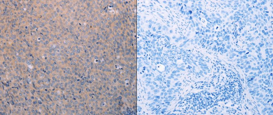
Predicted cell location: Cytoplasm. Positive control: Human cervical and thyroid cancer tissue. Recommended dilution: 1/25-100 The image on the left is immunohistochemistry of paraffin-embedded human cervical cancer tissue using ANKRD26 antibody at dilution 1/25, on the right is treated with the synthetic peptide. (Original magnification:x 200)
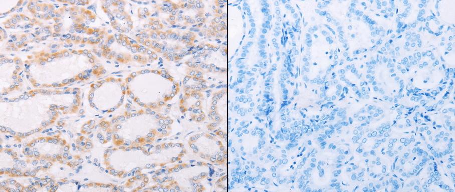
Predicted cell location: Cytoplasm. Positive control: Human cervical and thyroid cancer tissue. Recommended dilution: 1/25-100 The image on the left is immunohistochemistry of paraffin-embedded human thyroid cancer tissue using ANKRD26 antibody at dilution 1/25, on the right is treated with the synthetic peptide. (Original magnification:x200)
Primary Antibodies
| Catalog No. | Host | Iso. | Clone | Pres. | React. | Applications | |
|---|---|---|---|---|---|---|---|
| TA315188 | ANKRD26 antibody |
||||||
| Rabbit polyclonal anti-ANKRD26 antibody | |||||||
 |
Rabbit | IgG | Purified | Hu | E, P |
0.1 ml /
$335.00
|
|
| OriGene Technologies, Inc. | |||||||
| TA319345 | ANKRD26 (1-15) antibody |
||||||
| Rabbit polyclonal anti-Ankrd26 antibody | |||||||
 |
Rabbit | IgG | Aff - Purified | Bov, Hu, Ms, Rt | E, WB |
0.1 mg /
$515.00
|
|
| OriGene Technologies, Inc. | |||||||
| TA324215 | ANKRD26 antibody |
||||||
| Anti-ANKRD26 Rabbit Polyclonal Antibody | |||||||
 |
Rabbit | IgG | Purified | Hu, Ms, Rt | E, P |
0.1 ml /
$335.00
|
|
| OriGene Technologies, Inc. | |||||||
 +1 additional image +1 additional image |
|||||||