AKR1C4 antibody
Principal name
AKR1C4 antibody
Alternative names for AKR1C4 antibody
Chlordecone reductase, CDR, CHDR, 3-alpha-HSD1, Dihydrodiol dehydrogenase 4, DD4, DD-4, HAKRA, Aldo-keto reductase family 1 member C4, 3-alpha-hydroxysteroid dehydrogenase type I
SwissProt ID
P17516 (Human)
Gene ID
1109 (AKR1C4)
Ncbi ID
Available reactivities
Available hosts
Available applications
Enzyme Immunoassay (E), Immunocytochemistry/Immunofluorescence (ICC/IF), Western blot / Immunoblot (WB), Paraffin Sections (P), ELISA (detection) (E(detection)), Immunoprecipitation (IP)
Background of AKR1C4 antibody
- Kit Component:
- - KN204731G1, AKR1C4 gRNA vector 1 in pCas-Guide vector
- KN204731G2, AKR1C4 gRNA vector 2 in pCas-Guide vector
- KN204731D, donor vector containing left and right homologous arms and Luciferase-Puro functional cassette
- GE100003, scramble sequence in pCas-Guide vector - CRISPR Manual:
- CRISPR/CAS9 Genome Editing Manual
General readings
Stayrook KR. et al. (2008) Mol Pharmacol. 73:607-612.
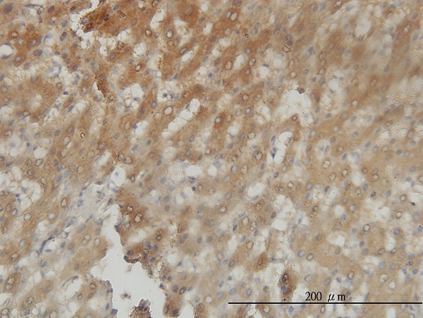
Immunoperoxidase of monoclonal antibody to AKR1C4 on formalin-fixed paraffin-embedded human liver tissue. [antibody concentration 3ug/ml]

Immunofluorescence of monoclonal antibody to AKR1C4 on HepG2 cell. [antibody concentration 15 ug/ml]
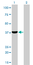
Western Blot analysis of AKR1C4 expression in transfected 293T cell line by AKR1C4 monoclonal antibody (M01), clone 2C11.Lane 1: AKR1C4 transfected lysate(37.2 KDa).Lane 2: Non-transfected lysate.
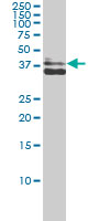
AKR1C4 monoclonal antibody (M01), clone 2C11 Western Blot analysis of AKR1C4 expression in MCF-7 ( Cat # L046V1 ).
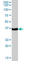
AKR1C4 polyclonal antibody ( Cat # PAB7023 ) ( 0.1 µg/ml ) staining of human liver lysate ( 35 µg protein in RIPA buffer ) . Primary incubation was 1 hour. Detected by chemiluminescence.
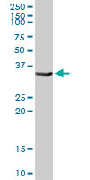
AKR1C4 MaxPab rabbit polyclonal antibody. Western Blot analysis of AKR1C4 expression in human liver.
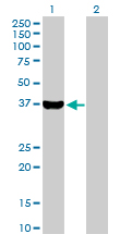
Western Blot analysis of AKR1C4 expression in transfected 293T cell line (H00001109-T01) by AKR1C4 MaxPab polyclonal antibody.Lane 1: AKR1C4 transfected lysate(37.20 KDa).Lane 2: Non-transfected lysate.
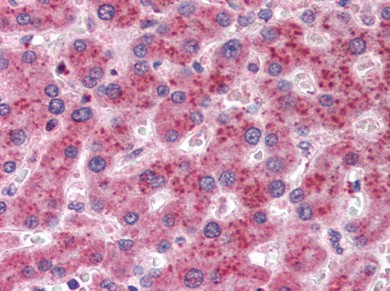
Liver: Formalin-Fixed, Paraffin-Embedded (FFPE)
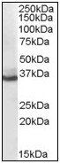
Antibody (0.1 µg/ml) staining of human liver lysate (35 µg protein in RIPA buffer). Primary incubation was 1 hour. Detected by chemiluminescence.
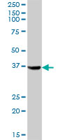
AKR1C4 MaxPab polyclonal antibody. Western Blot analysis of AKR1C4 expression in human liver.
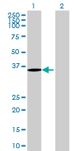
Western Blot analysis of AKR1C4 expression in transfected 293T cell line (H00001109-T01) by AKR1C4 MaxPab polyclonal antibody.Lane 1: AKR1C4 transfected lysate(35.53 KDa).Lane 2: Non-transfected lysate.
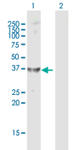
Western Blot analysis of AKR1C4 expression in transfected 293T cell line (H00001109-T01) by AKR1C4 MaxPab polyclonal antibody.Lane 1: AKR1C4 transfected lysate(37.20 KDa).Lane 2: Non-transfected lysate.
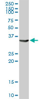
AKR1C4 MaxPab rabbit polyclonal antibody. Western Blot analysis of AKR1C4 expression in human liver.
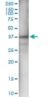
Immunoprecipitation of AKR1C4 transfected lysate using anti-AKR1C4 MaxPab rabbit polyclonal antibody and Protein A Magnetic Bead (U0007), and immunoblotted with AKR1C4 MaxPab mouse polyclonal antibody (B01) (H00001109-B01).
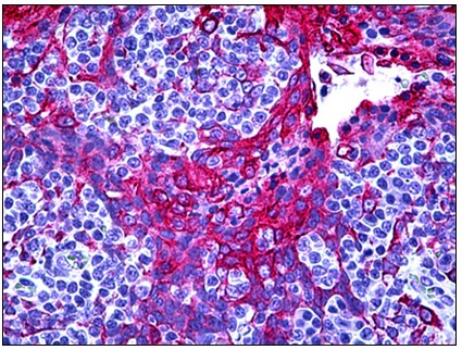
Human Tonsil, Squamous Epithelium: Formalin-Fixed, Paraffin-Embedded (FFPE)
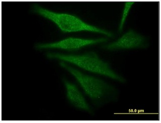
Immunofluorescence of monoclonal antibody to AKR1C4 on HepG2 cell. [antibody concentration 15 µg/ml]
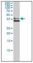
AKR1C4 monoclonal antibody, clone 2C11 Western Blot analysis of AKR1C4 expression in MCF-7.
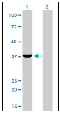
Western Blot analysis of AKR1C4 expression in transfected 293T cell line by AKR1C4 monoclonal antibody, clone 2C11.
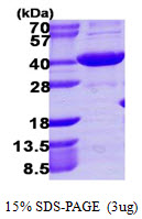

Recombinant human AKR1C4, 1-323aa, His-tagged
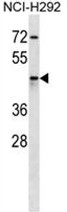
AKR1C4 Antibody (N-term) (Cat. #AP50134PU-N) western blot analysis in NCI-H292 cell line lysates (35 µg/lane).This demonstrates the AKR1C4 antibody detected the AKR1C4 protein (arrow).

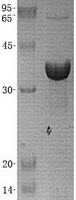

TA303107 (0.1µg/ml) staining of human liver lysate (35µg protein in RIPA buffer). Primary incubation was 1 hour. Detected by chemiluminescence.

Sample (30 ug of whole cell lysate). A: Hela. 10% SDS PAGE. HSD3a antibody. TA307983 diluted at 1:1000.
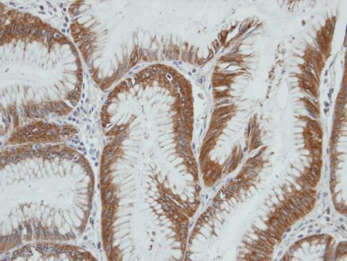
Immunohistochemical analysis of paraffin-embedded gastric ca, using AKR1C4(TA307983) antibody at 1:100 dilution.
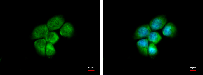
HSD3a antibody [N1C2] detects HSD3a protein at cytoplasm and nucleus by immunofluorescent analysis. Sample: A431 cells were fixed in 4% paraformaldehyde at RT for 15 min. Green: HSD3a protein stained by HSD3a antibody [N1C2] (TA307983) diluted at 1:500. B
Lysates
| Catalog No. | ||
|---|---|---|
| LY419725 | AKR1C4 overexpression lysate |
|
 |
0.1 mg /
$295.00
|
|
| OriGene Technologies, Inc. |