NAT1 antibody
Principal name
NAT1 antibody
Alternative names for NAT1 antibody
AAC, Arylamine N-acetyltransferase 1
Ncbi ID
9606, NP_000653, NP_000653.3, NM_000662, NM_001160179, NM_001160171, NP_032699.1, NM_001160175, NM_001160176, NM_001160170, NM_001160172, NM_001160173, NM_001160174
Available reactivities
Available hosts
Available applications
Enzyme Immunoassay (E), Western blot / Immunoblot (WB), ELISA (detection) (E(detection)), Immunoprecipitation (IP), Paraffin Sections (P)
Background of NAT1 antibody
- Kit Component:
- - KN310742G1, Nat1 gRNA vector 1 in pCas-Guide vector
- KN310742G2, Nat1 gRNA vector 2 in pCas-Guide vector
- KN310742D, donor vector containing left and right homologous arms and Luciferase-Puro functional cassette
- GE100003, scramble sequence in pCas-Guide vector - CRISPR Manual:
- CRISPR/CAS9 Genome Editing Manual
General readings
Gu J., et al. (2005) Multat Res. 581(1-2):97-104.
Kiss I., et al. (2004) Anticancer Res. 24(6):3965-70.
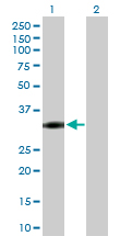
Western Blot analysis of NAT1 expression in transfected 293T cell line (H00000009-T02) by NAT1 MaxPab polyclonal antibody.Lane 1: NAT1 transfected lysate(33.90 KDa).Lane 2: Non-transfected lysate.
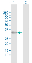
Western Blot analysis of NAT1 expression in transfected 293T cell line (H00000009-T02) by NAT1 MaxPab polyclonal antibody.Lane 1: NAT1 transfected lysate(33.90 KDa).Lane 2: Non-transfected lysate.
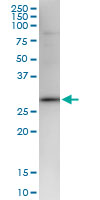
Immunoprecipitation of NAT1 transfected lysate using anti-NAT1 MaxPab rabbit polyclonal antibody and Protein A Magnetic Bead (U0007), and immunoblotted with NAT1 MaxPab mouse polyclonal antibody (B01) (H00000009-B01).
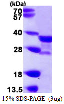

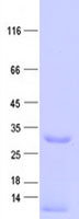
Recombinant human NAT1, 1-290 aa, His-tagged
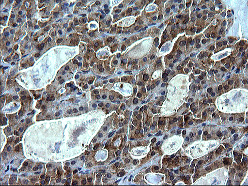
Immunohistochemical staining of paraffin-embedded Carcinoma of Human thyroid tissue using anti-NAT1 Rabbit Polyclonal antibody. (TA590782)
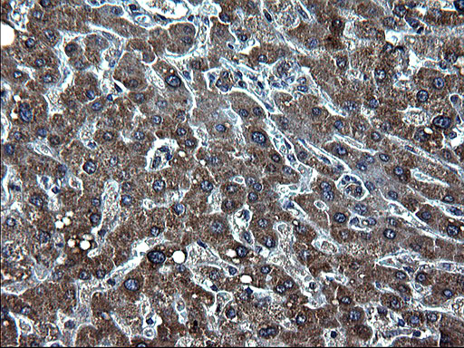
Immunohistochemical staining of paraffin-embedded Human liver tissue using anti-NAT1 Rabbit Polyclonal antibody. (TA590782)
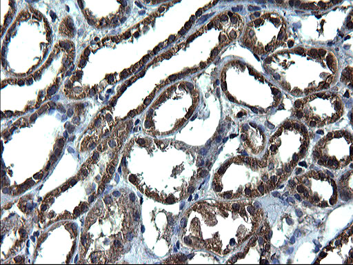
Immunohistochemical staining of paraffin-embedded Human Kidney tissue using anti-NAT1 Rabbit Polyclonal antibody. (TA590782)
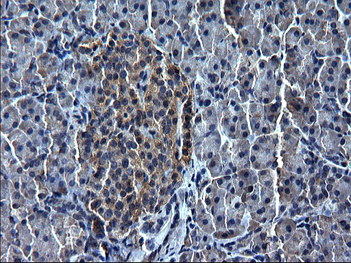
Immunohistochemical staining of paraffin-embedded Human pancreas tissue using anti-NAT1 Rabbit Polyclonal antibody. (TA590782)
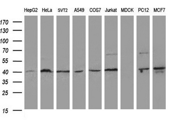
Western blot analysis of extracts (35ug) from 9 different cell lines by using anti-NAT1 polyclonal antibody (HepG2: human; HeLa: human; SVT2: mouse; A549: human; COS7: monkey; Jurkat: human; MDCK: canine; PC12: rat; MCF7: human).
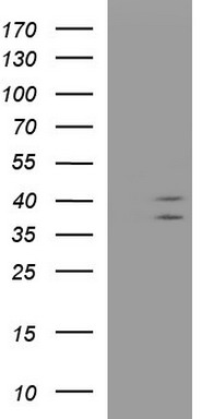
HEK293T cells were transfected with the pCMV6-ENTRY control (Left lane) or pCMV6-ENTRY NAT1 (RC221042, Right lane) cDNA for 48 hrs and lysed. Equivalent amounts of cell lysates (5 ug per lane) were separated by SDS-PAGE and immunoblotted with anti-NAT1.
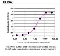
ELISA: NAT1 Antibody

Sample (30 ug of whole cell lysate). A: Hela. 12% SDS PAGE. TA308422 diluted at 1:1000.
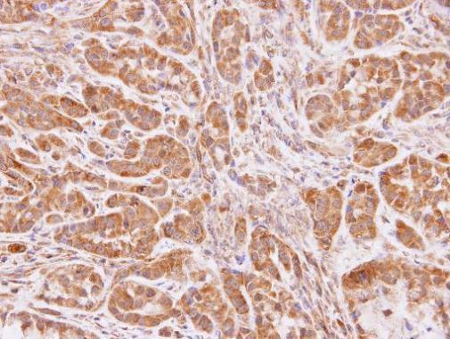
Immunohistochemical analysis of paraffin-embedded A549 xenograft, using NAT1(TA308422) antibody at 1:100 dilution.
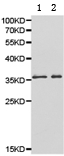
Predicted band size: 34 kDa . Positive control: A431 and HeLa cell lysate. Recommended dilution: 1/500-2000. (Gel: 10%SDS-PAGE Lane1: A431 cell lysate Lane2: HeLa cell lysate Lysates: 40 ug per lane Primary antibody: 1/500 dilution Secondary antibody: Goat anti Rabbit IgG - H&L (HRP) at 1/10000 dilution Exposure time: 1 minute)
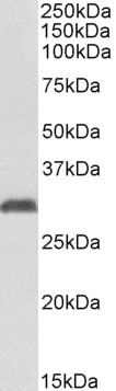
TA326332 (1µg/ml) staining of Human Erythrocytes lysate (35µg protein in RIPA buffer). Primary incubation was 1 hour. Detected by chemiluminescence.
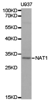
Western blot analysis of extracts of U937 cell line, using NAT1 antibody.
Primary Antibodies
| Catalog No. | Host | Iso. | Clone | Pres. | React. | Applications | |
|---|---|---|---|---|---|---|---|
| TA308422 | NAT1 antibody |
||||||
| Rabbit Polyclonal antibody to NAT1 (N-acetyltransferase 1 (arylamine N-acetyltransferase)) | |||||||
 |
Rabbit | IgG | Aff - Purified | Hu | P, WB |
0.1 ml /
$415.00
|
|
| OriGene Technologies, Inc. | |||||||
 +1 additional image +1 additional image |
|||||||
| TA326332 | NAT1 (C-term, 272-286) antibody |
||||||
| Goat Anti-NAT1 (aa272-286) Antibody | |||||||
 |
Goat | Aff - Purified | Hu | WB |
0.1 mg /
$360.00
|
||
| OriGene Technologies, Inc. | |||||||