Goat IgG antibody
Principal name
Goat IgG antibody
Alternative names for Goat IgG antibody
Goat Immunoglobulin G
Available reactivities
Available hosts
Available applications
Western blot / Immunoblot (WB), Enzyme Immunoassay (E), Frozen Sections (C), Immunocytochemistry/Immunofluorescence (ICC/IF), Flow Cytometry (F), Paraffin Sections (P), Immunodiffusion (ID)
General readings
J.A. Titus, P.P. Haugland, S.D. Sharrow, D.M. Segal J. Immunol. Methods 50; 193, 1982. (Conjugation)
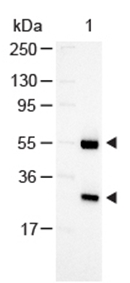
Western Blot of Rabbit anti-Goat Antibody Alkaline Phosphatase Conjugated:
Lane 1: Goat IgG, Load: 100 ng per lane Secondary antibody Alkaline Phosphatase Conjugated Rabbit Anti-Goat Antibody at 1/1000 for 60 min at RT
Block: MB-070 30 min RT
Predicted/Observed size: 55 and 28 kDa, 55 and 28 kDa
Lane 1: Goat IgG, Load: 100 ng per lane Secondary antibody Alkaline Phosphatase Conjugated Rabbit Anti-Goat Antibody at 1/1000 for 60 min at RT
Block: MB-070 30 min RT
Predicted/Observed size: 55 and 28 kDa, 55 and 28 kDa
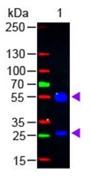
Western Blot of Rabbit anti-Goat IgG (H&L) Antibody Fluorescein Conjugated:
Lane 1: Goat IgG, Load: 50 ng per lane Secondary antibody: Goat IgG (H&L) Antibody Fluorescein Conjugated at 1/1,000 for 60 min at RT
Block: MB-070 for 30 min at RT
Predicted/Observed size: 55 and 28 kDa, 55 and 28 kDa
Lane 1: Goat IgG, Load: 50 ng per lane Secondary antibody: Goat IgG (H&L) Antibody Fluorescein Conjugated at 1/1,000 for 60 min at RT
Block: MB-070 for 30 min at RT
Predicted/Observed size: 55 and 28 kDa, 55 and 28 kDa
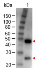
Western Blot of Rabbit anti-GOAT IgG (H&L) Antibody Peroxidase Conjugated:
Lane 1: Goat IgG, Load: 100 ng per lane Secondary antibody GOAT IgG (H&L) Antibody Peroxidase Conjugated at 1/1,000 for 60 min at RT
Block: MB-070 for 30 min at RT
Lane 1: Goat IgG, Load: 100 ng per lane Secondary antibody GOAT IgG (H&L) Antibody Peroxidase Conjugated at 1/1,000 for 60 min at RT
Block: MB-070 for 30 min at RT
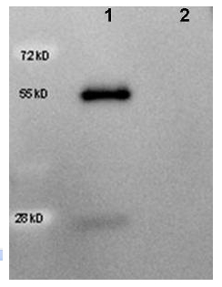
Western Blot of HRP conjugated anti-Goat IgG antibody R1318HRP showing detection of 50ng of Goat IgG (Lane 1) but not Human IgG (Lane 2). Samples were separated by 4-20% SDS-PAGE under reducing conditions and transferred to nitrocellulose membrane. The blot was blocked overnight at 4°C in 5% BSA in TBS. A 1/5,000 dilution of antibody in Blocking Buffer for Fluorescent Western Blotting was used to probe the membrane at RT for 1 h. The image was developed using Chemiluminescent FemtoMax™ Super Sensitive HRP Substrate (p/n Femtomax-020) for one minute.
Secondary Antibodies
| Catalog No. | Host | Iso. | Clone | Pres. | React. | Applications | |
|---|---|---|---|---|---|---|---|
| R1317B | Goat IgG (H+L chain) |
||||||
 |
Rabbit | Biotin | C, E, WB |
2 mg /
$220.00
|
|||
| OriGene Technologies GmbH | |||||||